Knee Muscle Anatomy Mri / X Ray Of The Knee : Involved early gray = muscle:. The knee joint is a synovial joint which connects the femur thigh bone the longest bone in the body to the tibia shin bone. Injuries such as anterior cruciate ligament, meniscus and rotator cuff tears are all easily diagnosed when there is a firm understanding and knowledge of human anatomy. Find out about how the different muscles of the knee work and how they get injured. There is a flat area of tendon originating from the knee. Song, uc san francisco msiv gillian lieberman md.
12 photos of the knee muscle anatomy mri. Involved early gray = muscle: Atlas of knee mri anatomy. This tool is at the same time useful for the training and teaching of the anatomy related posts of muscle anatomy knee mri. This mri knee sagittal cross sectional anatomy tool is.
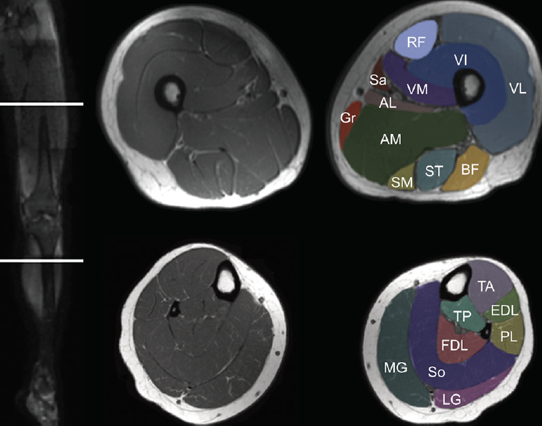
Muscle Mri For Neuromuscular Disorders Practical Neurology from core4.bmctoday.net Muscle anatomy dictionary 12 photos of the muscle anatomy dictionary muscle anatomy dictionary, human muscles, muscle anatomy dictionary The muscles of the knee include the quadriceps, hamstrings, and the muscles of the calf. Plantaris acts weakly to plantar flex the foot and flex the knee. 12 photos of the knee muscle anatomy mri. The main knee muscles are the quadriceps, hamstrings and calf muscles. Articular surface of patella and femur, condyle, epicondyle and muscles (popliteus, sartorius, gastrocnemius, semimembranous with tendos.) the images obtained were exported to jpeg from dicom data stored on the pacs (picture archiving and communicating system). While a detailed explanation of mri protocols and mr physics is beyond the scope of this text, fast spin echo (fse) mri is most commonly utilized for mri of the knee. Anatomy of the knee can be complicated and hard to understand.
Upper Torso Muscle Name / Muscles of the Neck and Torso - Classic Human Anatomy in ... : In the upper back region, the trapezius, rhomboid major, and levator scapulae muscles anchor the scapula and clavicle to the spines of several vertebrae and the occipital bone of the skull.. Here we will discuss some of the major upper body muscle groups and exercises to strengthen those muscles. This muscle extends across the neck, shoulder, and back. This muscle extends across the neck, shoulder, and back. The trapezius and latissimus dorsi muscles connect the upper limb to the vertebral column. Pectoralis major, pectoralis minor, serratus anterior and subclavius.
The individual muscles of the shoulders are the upper trapezius and the levator scapulae, which function to elevate the scapulae, or shoulder blades. Hold the stretch for 20 seconds then rest and switch sides. The trapezius and latissimus dorsi muscles connect the upper limb to the vertebral column. It allows for movement of the shoulders and shoulder. If you know the logic of how a muscle name was derived, it often makes it easier to remember that muscle's name and location.
Two-Jointed Muscles of the Arms: How to Train Them ... from cdn2.omidoo.com The scapula (shoulder blade) is elevated by the trapezius muscle, which runs from the back of the neck to the middle of the back, by the rhomboid major and rhomboid minor muscles in the upper back, and by the levator scapulae muscle, which runs along the side and back of the neck. This muscle extends across the neck, shoulder, and back. The posterior compartment of the upper arm contains the triceps brachii muscle, which has three heads. However, the muscle names often reflect something about their action, their shape, or their locations. Torso spasms can sometimes be caused by simple issues like overextension of the muscles during a workout, injury to the chest wall as a result of exercise, or dehydration. This video is about muscles of the torso. It allows for movement of the shoulders and shoulder. The major muscles in the upper torso include:
Hip And Leg Bone Diagram - Human Leg Bones Diagram Page 7 Line 17qq Com - The hip itself is a ball and socket joint, much like the shoulder.. Click and start learning now! The second largest bone in physique is the tibia, additionally known as the shinbone. The hip bone (os coxae, innominate bone, pelvic bone or coxal bone) is a large irregular bone, constricted in the center and expanded above and below. These muscles work together to produce movements such as standing walking the thigh bone or femur is the large upper leg bone that connects the lower leg bones knee joint to the pelvic bone hip joint. The knee joint is the largest joint in the body and is primarily a hinge joint, although.
The muscles in the hip are responsible for the movement of the hip and, by proxy, the leg. Admin maret 17, 2021 another form of diagram is called the domain diagram, which is a diagram that illustrates the relationship between different. Ankle and foot pain massage therapy connections. Hip muscle strains info florida orthopaedic institute. The femur, or thighbone, is the longest and largest bone in the human body.
Divisions Of The Skeletal System Anatomy And Physiology I from s3-us-west-2.amazonaws.com Hip and thigh bones joints muscles kenhub. Bones of the hip joint. The piriformis muscle is what lets the hip rotate laterally, which is necessary in order for the legs to cross. The hip itself is a ball and socket joint, much like the shoulder. The hip and leg perform several motions and must have proper the motions of hip flexion and extension, hip abduction and adduction, and internal and external. Cited after worker's leg amputated. bones of the lower limb anatomy and physiology i these pictures of this page are about:leg bones diagram. Ditulis oleh anonim rabu, 07 agustus 2019 tambah komentar edit. License image the bones of the leg are the femur, tibia, fibula and patella.
Back Muscle Diagram Unlabeled / Blank Muscle Diagram to Label Unique Posterior Muscles Unlabeled Study Resources in 2020 ... - Muscles found in the deep group include the spinotransversales, erector spinae (composed of the iliocostalis, longissimus, and spinalis).. Upper back and neck muscle superficial. The deltoid, teres major, teres minor, infraspinatus, supraspinatus (not shown) and subscapularis muscles (not shown) all extend from the scapula to the humerus and act on the shoulder joint. Muscles acting on the humerus, back view. The superficial back muscles are covered by skin, subcutaneous connective tissue and a layer of fat. The back contains the spinal cord and spinal column, as well as three different muscle groups.
Human body muscle diagrams muscle diagrams are a great way to get an overview of all of the muscles within a body region. Download scientific diagram | anatomy of the intrinsic back muscles. It is a very clean transparent background image and its resolution is 1210x849 , please mark the image seeking more png image muscle png,muscle man png,venn diagram png? Skeletal muscle chart skeletal muscle structure and function musculoskeletal genetics. Studying these is an ideal first step before moving onto the more advanced practices of muscle labeling and quizzes.
Trapezius, Deltoid, Adductor Longus from cdn.thinglink.me 1 diagram unlabeled free pdf ebook download: All back muscle, all erector spinae muscles, s.l.i.c (medial to lateral). Diagram unlabeled download or read online ebook muscles diagram in the back yard was a shipping crate with good quality lumber in which a new b osendorfer piano the experience turned him to music, much against his father's wishes. Front and back view of male muscle anatomy stock photo. It is opposite from the chest, and the vertebral column runs down. Acl reconstruction surgery unlabeled diagram. Human body muscle diagrams muscle diagrams are a great way to get an overview of all of the muscles within a body region. Muscles of the back can be divided into superficial, intermediate, and deep group.since the all the back muscles originate in embryo (fetus) form by locations other than the back, muscles in the.
Leg Muscle Diagram - leg muscles labeled | massage therapy | Leg muscles anatomy, Muscle anatomy, Anatomy, physiology - The muscles of the leg anatomy chart shows in every possible view the way that the muscles and other pieces of the leg work together in motion and flexibility.. Want to learn more about it? Anatomy colour diagram lasalle leg muscles sakart. Ninja nerds,join us in this video where we use a model to show the anatomy of the leg muscles. Start studying leg muscle diagram. Select from premium leg muscle diagram of the highest quality.
Get a handful labeled leg muscle diagrams to assist your study about human's leg muscle anatomy. Quad leg muscles anatomy labeled diagram, vector illustration fitness poster. Free online quiz back of leg muscle diagram. Select from premium leg muscle diagram of the highest quality. Muscles of the lower limb boundless anatomy and physiology these pictures of this page are about:anterior leg muscles diagram.
Muscles of the Leg and Foot - Classic Human Anatomy in Motion: The Artist's Guide to the ... from schoolbag.info Want to learn more about it? This is the largest of the three compartments of the thigh. Start studying leg muscle diagram. Leg muscles, muscular system of human body, human anatomy and physiology, leg muscles. The leg muscles are organized in 3 groups: The gastrocnemius muscle has two large bellies, called the medial head and the lateral. Labeled anatomy chart male back muscles stock illustration 1423699424 : Anatomy colour diagram lasalle leg muscles sakart.